
Prostate cancer is one of the most common types of cancer and may cause serious problems if it spreads to other parts of the body. Although most men have localised propstate cancer, in some cases it can spread. Common symptoms may include blood in your urine or semen, pain or burning during urination, a weak or hard to start urine stream and feeling the sudden urge to urinate right away.
A PSMA PET scan is a nuclear medicine imaging technique that provides a clear picture of what patients are dealing with. It uses a radiotracer drug (68Ga-PSMA or 18F-PSMA) which is given to patients one hour prior to the examination. This drug binds specifically to PSMA proteins overexpressed in prostate cancer cells. The tracer bound to cancer cells can then be easily visualised using a PET scan. Following the scan, highly detailed images of the prostate gland and surrounding tissues could be obtained.
In contrast to other conventional scans, this scan is vital in evaluating and planning for the treatment. These scans show a high level of sensitivity in finding prostate cancer lesions, even at low concentrations. They allow finding out the exact location and size of tumours along with the information whether the cancer has spread to nearby lymph nodes or other organs. They are also a promising method in staging prostate cancer, which allows doctors to select the best treatment options. These scans are also used for evaluating cancer in bones and other soft tissues. They are helpful in finding out the recurrence of prostate cancer after initial treatment. Overall, it removes the potential for surgical biopies.
How the PSMA PET works
Prior to the scan, the patients are typically advised to fast for a few hours and stay properly hydrated. Upon arrival at the diagnostic facility, a nuclear medicine expert administers an injection through a vein in the arm. It contains a radioactive tracer, which travels through the body for nearly 70-80 minutes to distribute before the imaging process starts. It comes under the waiting period. The tracer is generally well tolerated and has minimal side effects.
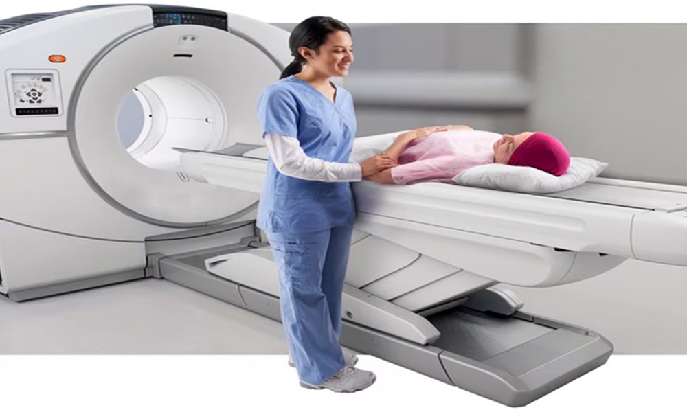
After the injection, the tracer circulates throughout the body and binds to any cancer cells that express PSMA. The patient is then asked to rest quietly. Once the uptake period is over, the patient is then needed to go into the scanning room. The scan usually lasts for 20-40 minutes.
Following the scan the patient can resume their normal activities and drink fluids to help excrete the tracer. The results offer detailed insights, helping clinicians to pinpoint the exact location, size, and spread of the disease with high accuracy.
Why this scan matters
This scan holds importance for its ability to spot prostate cancer, often with great detail. They are helpful in situations such as:
- Detecting a return of cancer when PSA levels begin to rise after treatment
- Before the treatment begins, it helps evaluate how far the condition has progressed
- It aids in tracking how well the ongoing therapies are working
- It aids clinical decisions which are related to surgery or radiation treatment
With sharper images as compared to other conventional methods, the PSMA PET CT scan plays a vital role in managing prostate cancer effectively.
Disclaimer: This information is for general awareness only and should not be considered as professional medical advice. Always consult a qualified healthcare provider for personalised guidance.